
Assess
Manage
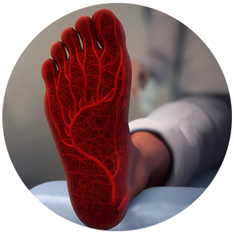
Preserve
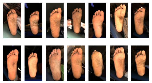
Assess
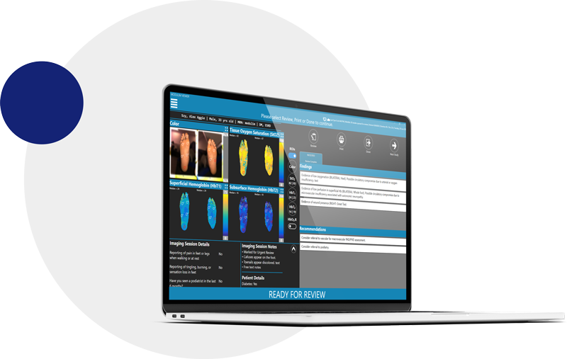
Assess patients at the point of care using tissue health biomarkers to help risk stratify, support care pathway decisions, and follow up frequency
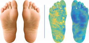
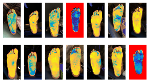
Physical exam showed no pathology. SFDI showed asymmetry, potential compromised circulation in left foot. Further vascular consultation may be needed.**
**High resolution recreations of data.
Manage
Manage patients with real time guidance by assessing the responsiveness to therapeutic interventions (e.g. targeted offloading, debridement, HBOT, stent patency, etc.)
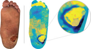
Reduced perfusion in periwound area identified. Further intervention (e.g. debridement) may be needed to aid in healing.**
**High resolution recreations of data.
Preserve
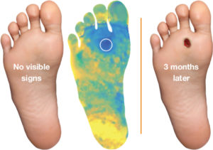
Identify focal (pre-ulcer) and systemic circulatory issues sooner allowing earlier intervention
Physical exam showed no pathology. SFDI showed focal area of compromised circulation at second metatarsal head. No intervention performed, 3 months later patient returns with ulceration (Mazhar et al., 2019).**
**High resolution recreations of data.